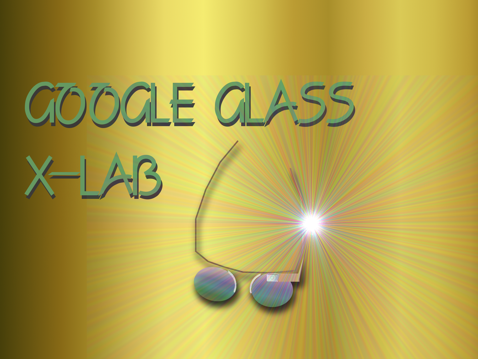
Keratoconus | Eye Doctors Contact Lenses and New Options for Treatment

Keratoconus Courtesy of Indiana University Department of Ophthalmology

Keratoconus is an eye disease that causes vision to gradually worsen over time, as the transparent corneal tissue that covers the front of the eye thins and bulges forward, forming the cone shape that keratoconus is named for. Rapid increases in nearsightedness and astigmatism are common with frequent changes in your eyeglass prescription. Scarring of the cornea can also occur resulting in significant vision loss.

Keratoconus Symptoms Your Optometrist may discuss

  • The appearance of long linear light streaks in your eyesight at night
  • Visual Glare and halos around lights, especially car headlights and taillights at night
  • Double Vision
  • Distorted Vision
  • Blurry Vision making it difficult to read
  • Ghost like eye images of white light surrounding objects you are viewing, sometimes noticed as multiple dots when viewing on small light image-can be seen with one eye
  • Eye Sensitivity to light
  • Eye Doctors Agree Keratoconus usually show up most commonly between the teenage years of 16 up to age 30

When this corneal eye disease manifests at earlier ages optometrists often find a more aggressive form with ongoing, frequent changes in your eyeglass or contact lens prescription. The best guess is the occurrence rate is about 1 in 2000 people. It is hard for eye doctors to pin down the exact rate of this eye disease because it can be very mild and remain undiagnosed, especially when it burns out early (form fruste), or stops progressing after several years. Corneal Keratoconus creates irregular astigmatism, causing curvatures on the cornea tissue that are not nice smooth curves. Instead the eye curvature resembles the surface of a potato with dips and valleys on a very irregular shape. This type of shape makes it very difficult for your optometrist to prescribe an eye prescription for eyeglasses that results in clear vision. The lenses would have to be made in very strange shapes to result in clear vision. Even lenses that have been designed in this manner are rendered ineffective the moment your eyes look off the center of the lens. Irregular astigmatism also occurs without kerataconus, but it does not tend to progress and result in the characteristic steepening of the cornea resembling a cone shaped area. Usually kerataconus presents in one eye and over time well over half of the eye patients will have both eyes involved.

The cause of Kerataconus and the changes in the cornea are surprisingly not well known by the optometric research clinics at this time. In the past, much speculation centered around eye rubbing and ocular allergies. Some eye physicians have speculated that Keratoconus is triggered by eye rubbing that starts an inflammatory cascade in the cornea. Frequent eye rubbing also could cause mechanical tissue breakdown in areas of the cornea that are already compromised. Research by eye doctors has shown between 6-8% of patients with kerataconus have a family history, indicating there is a genetic component in some cases. Several areas of chromosomes have been identified as potential genetic markers and are being investigated further. Also, certain eye diseases such as retinitis pigmentosa, retinopathy of prematurely (damage to the retina tissue in the back of the eye from premature birth), Leber’s congenital amaurosis (a degenerative disease of the optic nerve), and vernal keratoconjunctivitis (a type of allergic eye disease which creates itchy eyes and frequent) appear to have some correlation. Some disease of the body also have a degree of co-occurrence with Keratoconus- Ehlers-Danlos syndrome, Down syndrome, osteogenesis imperfecta, pseuodoxanthoma elasticum), mitral valve prolapse in the heart, Laurence-Moon-Biedl syndrome, Rieger’s syndrome and neurofibromatosis. Several of these diseases interfere with normal collagen development and may precipitate kerataconus by disrupting collagen development in the cornea.

The changes to the cornea from Keratoconus are mostly unknown. The cornea consists of 5 layers and is about 1/2 mm thick (550 microns or about the width of 5 human hairs). The epithelium layer is the surface layers of cells. Underneath the eyes epithelium layers is a thin basement membrane sitting on the anterior limiting membrane, also know as bowman’s membrane. The bulk of the corneal thickness is in the stromal layer, where the collagen protein fibers run across the cornea, adding the tensile strength. Tensile strength is the degree a material can be stressed and still return to it’s original state and shape. Collagen is the memory material of the cornea. The structure of collagen changes in the center area of the cornea with shorter fibers, that cross more, run at different angles, run though each other, form connections to Bowmans membrane, and also form connections originating from Bowman’s membrane. It has been suggested from research by Jan P.G. Bergmanson, OD, PhD, PhD h.c, DSc, & Jessica H. Mathew, OD that this alteration in structure near the central cornea may help explain the nature of Keratoconus in the future, With shorter fibers running in differing directions with various connections the central cornea would seem to be more prone to breakdown of the normal collagen structure. Optometrists have found the bulging cone area characteristic of Keratoconus cones usually form close to the central cornea, slightly inferior, which seems to substantiate the altered central corneal tissues may play a part in the eye condition. Early changes may occur in the surface epithelial cells disrupting the basement membrane. When keratoconus begins, whatever the cause may be, enzymes increase and start damaging the epithelial basement membrane. This is the membrane formed underneath the lowest level of epithelial cells. Subsequent breaks in the corneal anterior limiting membrane occur and the cornea starts to thin centrally, probably due to the susceptibility of the different surface anatomy of the collagen fibers under lying Bowman’s membrane. As these breaks occur the surface epithelial cells can contact the stromal level of the cornea where most of the structural framework of this eye tissue is located. Small proteins called cytokines are released and alter the fluids around the cells, leading to scarring of the cornea.  Stromal fibers may move through the anterior limiting membrane. Whatever the cause, a disruption of the normal collagen structure causes the memory shape to lose its capacity and irregular shaped corneas to subsequently develop. There are indications of changes in the different enzymes that degrade proteins and induce changes in the collagen and the spaces surrounding the cells in the cornea. Cathepsins are one type of protein that increase as kerataconus starts to occur. These could lead to destruction of the so called extra cellular matrix, the substances surrounding the cells and lead to degenerative effects in the cornea.
They may also indirectly cause a reduction in the antioxidants and increase oxidative damage to the cornea, another theory that has been proposed. Matrix metalloproteinase-2 is also activated and changes the extra cellular matrix surrounding the corneal cells. Keratocytes are numerous cells in the cornea that produce the collagen for the fibers and the extracllular matrix components, turning mostly dormant by birth. In Keratoconus they have been observed to have increased apoptosis (increased programmed cellular death). There us a reduction in the number of collagen fiber and they also reduce in diameter. Most likely, keratoconus will be found to be several different disease processes and also multifactoral. Multifactoral eye diseases have multiple factors that combine to create the eye condition. For instance, the different collagen structure in the central cornea makes the eye susceptible, changes in enzymes may alter the tissues and start causing minor breakdowns in the epithelial surface cells, enzyme changes may lead to increased oxidative stress further weakening the eye tissue, and constant rubbing of the eyes may push the eyes over the edge by inducing mechanical damage to the eye tissues that could only occur with a compromised cornea. A genetic alteration of the cornea could make the cornea of the eye more susceptible to the entire chain of events.

Your eye doctor will initially treat Keratoconus with contact lenses

Treatment of Keratoconus usually begins with a rigid gas permeable contact lens when vision can no longer be maintained clearly with spectacle lenses. Sometimes, a gas permeable lens will be fit over the top of a soft contact lens in a piggyback contact lens fitting, with a soft contact lens and a rigid gas permeable contact lens on top of it. While used years ago, piggyback contact lens fittings fell out of favor due to the complications from reduced oxygen flow with older soft contact lenses. With the new super oxygen permeable silicone hydrogel soft contact lenses, it is enjoying a small resurgence. It is primarily used to increase eye comfort for the keratoconic eye patient. There are also combination contact lenses available today, such as the SynergEyes contact lens that is a rigid contact lens with a soft skirt attached surrounding the lens. The primary issue with Keratoconic contact lens fittings is matching the steeply curved cone with the surrounding flatter eye tissue, while dealing with the irregularities of curvature that are present. While custom mapping technology is highly touted as the way to achieve the required fit, the truth is observation of contact lenses on a keratoconic eye by an optometrist and adjustments based on how dyes accumulate under the contact lenses is still the most accurate method to achieve an excellent final fit. Due to the drastic changes in curvature the contact lenses require multiple different curves as you move toward the edge of the lens. While many different lenses have been developed with special names as the ultimate Keratoconus contact lens, they are all variations on the basic concept of a steep contact lens center and a gradient of changing curvatures to the edge. Rigid contact lenses work because the light bending capacity of the tears is very close to the light bending capacity of the cornea. The tears fill in between the irregular eye surface and the smooth surface on the back of the lens. This essentially removes the irregular astigmatism and nearsightedness by utilizing the back contact lens surface as a new regular surface where light is altered, and often restores the corrected eyesight close to 20/20. Eye glasses may only achieve 20/40 vision or much worse because the irregular surface remains. Occasionally Scleral rigid gas permeable lenses are used. These are gas permeable lenses larger than normal that extend out onto the white part of the eye. All contact lenses today are gas permeable, or designed to let air pass through to keep the cornea healthy. Soft contact lenses are usually not referred to as gas permeable because of historical changes. Hard lenses were the first contact lenses and they were made of a material that passed no oxygen through the lens. When changes were made to the polymers used to make hard contact lenses that allowed them to breathe or pass needed air to the underlying cornea, they were renamed rigid gas permeable contact lenses. Rigid because they are still a hard material with only 1-2% water, and gas permeable because unlike the older hard contact lenses they now transmitted air to the eyes. Soon they became called by the acronym of RGP’s to save a few words (even prior to the texting era). With time they also came to be referred to as gas perms, in spite of the fact that all soft contact lenses are also gas permeable. Soft contact lenses are never rigid however, as they normally are composed of about 50% water. This softness comes at the price of increased flexibility and they drape across the eye, imitating the irregularities of a Keratoconic cornea and do not correct the vision back to optimum levels. Once your eye doctor has achieved an excellent fit and optimized your contact lens prescription, there may be frequent changes in your contact lens prescription as the Keratoconus goes through progressive changes.

Your Eye Doctor May Be Able to Avoid Corneal Transplants

While only 10-20% of eyes will undergo ongoing serious changes, they do present challenges to fitting contact lenses on eyes with Keratoconus. At some point, scarring of the cornea can start to occur and patients become intolerant to contact lenses. In years past, the only remaining option was a corneal transplant. While corneal transplants enjoy a relatively high success rate, there are still risks and problems. Recently there have been some new exciting options starting to evolve.

Permanent Contact Lenses-Intacs

Intacs are small rigid half rings similar to portions of a gas permeable contact lens that are implanted in the cornea. They were originally developed to reverse nearsightedness, but did not prove as effective as originally thought and were replaced by LASIK eye correction procedures. A few years ago they found a new use in stabilizing Kerataconus. They are not a cure for Keratoconus, but can restore some more regularity and allow some patients to continue contact lens wear while avoiding a corneal transplant. They also appear to have some effect in decreasing the rate of change in Keratoconus. While they are promoted as being completely removable and reversible if patients have problems, this is not entirely true. About 10-15% of Intacs cause some complications and issues that cannot be resolved if they are removed. Still, it is a better option than jumping straight to a corneal transplant.

Keratoconus Corneal collagen cross-linking therapy

Corneal collagen cross-linking therapy (CXL) is intended to stabilize the tissue by forming more bonds between the existing collagen fibers and also increasing the size of the fibers, making the cornea much firmer and less likely to continue deforming. It involves pretreatment the cornea with riboflavin (Vitamin B2) for 30 minutes then using radiation from the ultraviolet A band light spectrum (normally around 370 nanometers) to increase the cross links over about a 30 minute period. While it has been more extensively in Europe, it is starting to enter the U.S. market. The riboflavin acts to keep the UVA from completely passing through the cornea so the UV can act to create more cross links. Riboflavin also may have a photo reactive effect that further increases cross linking of the collagen bands. Questions still surround this treatment. It is not an FDA approved treatment in the United States but is undergoing clinical trials, and currently is used off label as a treatment for Keratoconus. The FDA (Food and Drug Administration) regulates drugs and medical devices but not doctors. Any procedure that uses drugs or medical devices can be performed by a doctor if you are properly informed, share in the decision, and it has an acceptable possibility of helping. The riboflavin still allows a significant amount of UVA to pass through the cornea. This could potentially increase future risks for cataract development. UVA with riboflavin is cytotoxic (damaging to cells) and can damage the endothelial cells that line the back of the cornea and are vital for its long term health. Corneal thickness needs to be factored to keep this type of damage far enough away from the endothelium cells of the cornea. A minimum corneal thickness of 400 microns has been suggested but a better choice would be 450 to 500 microns. Cellular damage to the keratocytes, changes to the matrix of the cornea, and changes to the epithelium do occur in the cornea after the procedure. Normally they regenerate over the next 6 months. Riboflavin has poor penetration into the cornea so the surface layers of epithelium cells need to be removed. It is unknown if the effect of increased rigidity created by this treatment will last indefinitely, or if there are any other long term problems from increasing the cross linking and rigidity of the cornea. Some cases of persistent haze, infections, and increased eye pressure reading have been noted. The increased eye pressures are presumably an artifact since we know thicker (more firm) corneas read artificially high with most current glaucoma instruments. With careful consideration about the stage of Keratoconus and treatment of the eye at the appropriate stage, cross linking of collagen fibers in the cornea appears to be a great addition to eye doctors armetarium in treating keratoconus. While it may improve the condition mildly in many patients, it should always be considered as a stabilizing treatment and not a curative treatment.

Future therapies will evolve. Cross linking collagen therapy is still in its infancy. Stem cell and genetic treatments may be seen at some time. Someday we will no longer be treating Keratoconus but acting to prevent it from ever distorting peoples vision and lives.

Optometrists in Fort Collins Usually Prescribe Prism Eyeglasses For Double Vision

Has your eye doctor prescribed prism eyeglasses for you or your child in a new eyeglass prescription? It could be due to crossed eyes, lazy eye, or some diseases of the eyes or body.

Prisms are thin pieces of the optical material that is used in prescription eyeglasses. You may remember them from high school physics. They have a base that is thicker and an apex that is thinner. Due to the light bending properties of the lens material, the thicker lens base slows light down as it passes through. Since the prism is thicker at the bottom base it slows light down longer than the top apex, and light changes direction down towards the base as it exits the prism. If the lens were equally thick throughout, it would slow the light down but the direction would remain unchanged. The amount of direction change is determined by the index of refraction of the material compared to the index of refraction of air. Lenses with higher indexes of refraction allow optometrists to make the ultra thin lenses that have drastically improved eye comfort and cosmetic appearances in recent years. The light bending capacity of a lens material is referred to as the index of refraction. The value in space in a vacuum is 1.0, essentially the same as in the earth’s atmosphere which is 1.0003.  Eye glass lenses now have indexes of refraction from 1.5 to 1.7, with the higher numbers being the thinnest.

Eyes that cross can turn in or out, and also one eye can turn up or down. Strabismus is the technical term for a turned eye. Eyes that turn out are referred to as exotropic while eyes that turn in are called esotropic. An eye that turns up is hypotropic and one that turns down is hypotropic. These are all considered as cases of strabismus, or eyes that actually do cross. Often they are mistakenly called “Lazy Eyes.”  A lazy eye is a different condition but it may result in a crossed eye. A combination of eyes turning laterally and vertically is common. When this results in double vision, it can be completely disruptive to mobility and lifestyle of the affected person. If the eye turns are present at birth, there may be no double vision present. The brain has the capacity to suppress or turn off the area of vision that results in the eyes perceiving double at early ages. When a person sees double, prisms, eye exercises known as vision therapy, and surgery are the three options eye doctors have to try and restore normal visual functioning. Prisms do not appear as a strange looking triangle in the lens. They usually show up as thicker and thinner edges on the eyeglass lenses. Normally they are ground into the shape of the lenses, but because of optical properties, some lenses can have the optical center repositioned to induce prism. There can be an adjustment to prism added to a prescription while the brain relearns how to interpret the eyes seeing single. When diseases such as strokes and diabetes cause double vision there can be some fluctuation over time, and frequent eyeglass prescriptions changes to adjust the amount of prism may be required. Temporary press on prisms known as Fresnel lenses may be used in some cases when change is expected but their optical quality tends to be very poor.

Your eye  doctor may prescribe prism in your eyeglasses for other reasons

Some people will only have a tendency for an eye to turn, and while it may not actually turn it will result in eyestrain, fatigue with reading, headaches from using your eyes, and other symptoms. These are referred to as heterophorias instead of strabismus. Instead of exotropia your optometrist would call it exophoria. Any term ending in phoria means a tendency to turn while tropia indicates an eye that is turned. To complicate matters, sometimes eyes will cross at one distance and not another, or when viewing in only certain fields of gaze. In cases of traumatic head injuries yoked prisms may be used to help retrain a disrupted visual system by altering spatial perception.

Some serious health problems can cause double vision, and any new onset or increasing condition of double vision should be thoroughly examined by your optometrist

When double vision occurs after the age of fifty, common causes are thyroid conditions, high blood pressure and diabetes. Try to notice when it occurs and what makes it worse to help your optometrist in treating your eye condition with the best methods possible. In the case of a brain tumor inducing double vision, a trip to the eye doctor could save your life.

Fort Collins Eye Doctors Birds And Amblyopia Treatment

Fort Collins Eye Doctors Birds And Amblyopia Treatment

If you have a child between the age of 8-27 with lazy eye (amblyopia), the National Eye Institute will soon be soliciting volunteers for a study on a new form of treatment involving the drugs levodopa and carbidopa. These drugs are precursors for the production of dopamine , one of the main neurotransmitters, and often are sued to treat Parkinson’s disease. Dopamine is synthesized in the retina tissue in the back of the eye in amacrine cells. When the eye is light adapted, a lazy eye will show reduced levels of dopamine production, Dopamine seems to have a number of roles, effecting contrast sensitivity (which is one form of vision reduced in a lazy eyes. Rescued levels also inhibit the eye elongating (or growing longer from front ot back). This action tends to make an eye farsighted with astigmatism (since it does not seem to effect chewnges in equitoriL growth).

Adding these precurseres to lazy eye treatment may help restore sight past the age where we expect good results. After age 7-8 success rates drop down to 20-30% at best. This study will compare levadopa with and without patching an eye to see if there is a marked improvement. It will be oral supplementation with Levadopa. The trial is not yet recruiting for subjects and will probably require travel out of state.

This is very noteworthy from the aspect of treating a neurodegenerative condition potentially with a pharmaceutical drug and maybe reversing sightloss at an age where recovery isn’t normally expected.

Free treatment for lazy eye? Well free eye care is worth what it costs-but a simple eye drop prescription  may be able to save you a thousand dollars or more in treatment fees. And that’s equivalent to a lot of free office visits.

Forget the pirate patch, eye drops are the treatment of choice today for lazy eye. No more daily struggles with your child to keep an eye patch on, one drop in the morning and your responsibility is done for the day. Lazy eye, (technically referred to as Amblyopia), is the most common cause of permanent sight loss in children and if not treated by age 7 to 9 it persists into adulthood  with permanent damage to the visual cortex area of the brain. It is the leading cause of vision loss in one eye in the 20-70 year old age group, occurring in around 3% of the population.

Most of the information on the success of treatment of lazy eye has been from limited,uncontrolled studies. There is a great opportunity for future learning about improving the treatment of lazy eye.


Amblyopia   is still usually treated with outdated methods of occlusion (patching) of the eye that sees well. Patching therapy typically has very poor compliance, especially since you cannot be with your child every minute of the day. Kids don’t like wearing patches after the novelty wears off and may be subjected to ridicule by peers. Evidence indicates that compliance is probably the most important factor in the outcome of treatment of lazy eye.


Using a cycloplegic eye drop that prevents the good eye from focusing forces the lazy eyes to work to see thing at closer distances. As a side effect of the drops the pupil of the good eye stays dilated, so it needs some form of UV protection in eyeglasses or sunglasses. This is not a new method but it has started catching on due to recent studies showing it is effective and very well accepted by parents and children. Without a patch blocking one eye completely the child is able to maintain some level of binocular vision which helps the treatment process. It also assures there is no impairment to peripheral vision, making it a safer alternative to a patch.

Atropine is the drop normally used but we substitute homatropine. It is safer and in my opinion there is really no need  to keep the effect lasting overnight while the child is asleep.

There is a tremendous cost and time saving to the parent due to less frequent office visits and a great reduction in the amount of eye exercises needed to restore the sight. If you think there is no way you can get a drop in your child’s eye every day we have a secret for that too. If your child has a lazy eye we can help make it easy and fun!

Welcome to Dr Kisling-Fort Collins Eye Doctors Choice Website. Our vision is to be the premier resource for answers about your eye symptoms and eye problems. You can use the search box to find answers to questions about your eyes. You will find articles on contact lenses, dry eyes, glaucoma, eye nutrition pinkeye ad other eye diseases.

There is also information about eye examinations, eyeglasses, prescription eye glasses for different conditions;nearsightedness,farsightedness, astigmatism, and presbyopia.

We carry a unique selection of eyeglasses for women, men and children with the latest lens technologies including glare free anti-reflection coatings, no line progressive 5th generation eye glass lens designs, transition sunglasses tints, thin and light hi-index prescription lenses.

We also try to make your vision insurance processing as simple as possible. We welcome most insurance including Vision Service Plan (VSP).

If there is a topic you would like more information on please feel free to leave a request in the comment box. Thank you for visiting our website and we hope we can become your partner in preventative eyecare for a lifetime of healthy eyesight!

Pseudotumor cerebri is an eye condition that can be confused with a brain tumor due to swelling of the optic nerve. Like a brain tumor, the fluid inside the skull that cushions the brain and flows around the spinal column develops elevated pressure.  It is also referred to as Idiopathic Intracranial Hypertension or Benign Intracranial Hypertension. It is more prevalent in  women between the ages of 20 and 50. Symptoms of pseudotumor cerebri are usually an undiagnosed chronic headache, and in more severe cases there may be visual symptoms such as double vision. Also nausea and vomiting may occur. There is an unusual effect of  pulsating sounds within the head that are synchronous with the heart rate referred to as   Pulse-synchronous tinnitus.

A routine eye exam is where the initial diagnosis of mild cases of pseudotumor cerebri is often first made. The optic nerve that enters the back of the eye appears elevated in both eyes, a condition referred to as papilledema. Because the nerve is an extension of the brain and enclosed by the same tissues and fluid that cushion the brain, elevation in the pressure inside the brain also cause the nerve to swell. Since most cases occur in young to middle age overweight females, an elevated nerve with a history of chronic headaches and otherwise healthy is often indicative of pseudotumour cerebri. Other visual symptoms include double vision, brief periods of blurred vision or dim vision, and occasionally patient will have temporary episodes of blindness in one or both eyes. Changes in posture such as suddenly standing up or bending over may elicit symptoms, as may coughing or sneezing.

Double vision may occur when looking to the side. This is caused by a defect in abduction or the ability of an eye to turn out. due to a restriction in the capacity for one or both eyes to turn out. This presents a confusing picture when taken out of context because it is not due to damage to the abducens nerve (6th Cranial Nerve)  that controls the eyes outward motion. In this case, the double vision is called a false localizing sign. Pain can occasionally be associated with eye movements.

Rarely, the pressure can become very high and cause severe problems with vision and alterations in  levels of awareness. Most of the time truly is benign idiopathic intracranial hypertension and is managed by weight loss and occasionally medications. Sometimes discontinuation of certain medication may resolve the problems. The most severe cases may require a shunt to drain the increased fluid from the ventricles in the brain into the abdominal cavity.

Traumatic Brain Injuries are common causes of undiagnosed vision symptoms. The brain is cushioned in a compartment but sudden changes in velocity can cause it to move slightly and cause axonal shearing (minute tearing of the nerves in the brain). People who have been in accidents, especially car accidents, often find their visual system seems disjointed days to weeks after the accident. They may have double vision, light sensitivity, poor focusing skills, difficulty reading, dry eyes, and a number of other eye problems. Even very low velocity accidents can result in trauma under the right circumstances.

Treatment is available. There are multiple modalities to treat dry eyes, special tints to reduce the photophobia (light sensitivity), special eyeglass corrections with prism to relieve double vision, and vision therapy (eye exercises) that can help alleviate other eye symptoms.

One of the outcomes of the war in Afghanistan and Iraq has been a large increase in eye injuries. Somewhere between 15-20% of evacuations have been due to eye injuries. Improved Body armor has led to survival of wounds that would have been lethal in the past but has left more veterans living with sight loss. There is currently increasing awareness in our military hospitals of traumatic brain injury eye problems.

Seat belts, helmets, and protective eyewear save lives and eyesight. Don’t be remiss in making sure your family is safe.

And this Memorial Day Holiday, consider a donation to the Veteran’s Plaza being planned for Spring Canyon Community in Fort Collins. Captain Diggs Brown is the visionary, planner, and fundraiser-see Veterans Plaza or Ray Martinez’s Commentary for more information on the Veterans Plaza in Sprng Canyon Park.

Be safe and see safe this holiday weekend!

A fun new movie I recently went to see was Monsters vs. Aliens. It started just before a wedding with a bride growing (not glowing) from meteorite exposure, to enormous height and strength. Reese Witherspoon supplies the voice. Not a great movie but it is fun and the technology is fascinating. Monsters vs. Aliens was released on almost every 3-D screen avaible today with a count over 2000 and growing.Projections are for 40 new 3-D films within the next three years. RealD technology utilizes circular polarization, a technique with the polarization varying in a clockwise or counterclockwise direction. In Optometrists offices, polarized lenses and images are utilized in one constant plane tests for lazy eye (amblyopia) or crossed eyes (strabismus). Eye Doctors also use polarized equipment when treating binocular dysfunction for eyestrain, double vision, and convergence insufficiency causing vision related reading problems. Quality sunglasses enhance vision by using a fixed plane polarization to block reflections that occur most frequently from the surface of water. The RealD technology is comes with a high price tag costing theatres $50,000 to $70,000 per screen for the digital projection equipment and additional licensing fees. That’s why there is a nice little surcharge ($2.50 in Fort Collins) for the special eyeglasses needed. The neat thing is the circular polarization helps you maintain binocular vision and lessens eyestrain when you tilt your head, unlike older technologies. Less squinting and your eyes won’t leave the theatre with dry eyes from staring throughout the film trying not to see double. The first 3-D film shown to a paying audience was The Power of Love in Los Angeles in 1922. It was projected with anaglyphs, which involves showing a green image to one eye and a red image to the other which are shown from slightly different view points. Anaglyphs are still used by Optometrist in vision therapy for problems like Computer Vision Syndrome. Looking to the future, Steven Spielberg started work on a 3-D system in 2005 using plasma screens that is supposed to work without any special glasses! Coming to a TV near you some day.